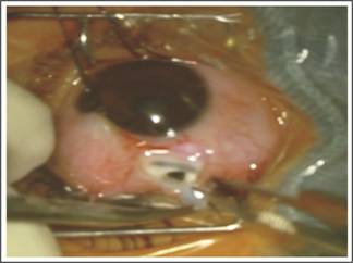
Original Article


Scleral Patch Graft in Spontaneous and Traumatic Corneoscleral Perforations


Sharjeel Sultan, Nisar A. Siyal, Nargis Nizam Ashraf, A. Rasheed Khokhar


Pak J Ophthalmol 2018, Vol. 34, No. 3

 . . . . . . . . . . . . . . . . . . . . . . . . . . . . . . . . . . . . . . . . . . . . . . . . . . . . . . . . . . . . .. . .. . . . . . . . . . . . . . . . . . . . . . . . . . . . . . . . . . .  . .

See end of article for

authors affiliations


…..………………………..


Correspondence to:

Sharjeel Sultan

Department of Ophthalmology Civil Hospital, Karachi

Email: sharj35@outlook.com


Purpose: To study the efficacy of scleral patch graft in spontaneous and traumatic corneoscleral perforations.

Study Design: interventional case series.

Place and duration of study: Civil hospital Karachi, unit 2 of ophthalmology, from March 2017 till August 2017.

Material and Methods: Patients underwent scleral patch graft in spontaneous and traumatic corneoscleral perforations. Convenience sampling was used for patient sampling. Baseline demographic characteristics such as age, gender and clinical parameters were included. Visual acuity (pre- and postoperative), details of surgery, final outcome and the complications were noted. Statistical package for Social Sciences (SPSS) version 20 was used for data analysis.

Results: Fifteen eyes of 15 patients were studied with scleral patch grafting in spontaneous and traumatic corneoscleral perforations. Ten patients (66.7%) were male and five (33.3%) were females. Age ranged from 10 to 80 years. Surgery was done in seven (46.7%) right eyes and eight (53.3%) left eyes. In five eyes (33.3%), postoperative visual acuity remained unchanged. Stable ocular surface was observed in nine patients (60.0%) and improvement was observed in one eye (case 3). In 3 patients (case 1, 10 and 12) keratoplasty was advised and one patient (case 13) was advised Evisceration.

Conclusion: Preserved scleral graft in spontaneous and traumatic corneoscleral perforations gives both functional and structural stability to eyes.

Keyword: Scleral patch graft, Corneoscleral perforations, Visual acuity.


Eye is a very delicate organ of the body so nature has protected it in a very hard bony socket. Injuries to eye are a common cause of emergency attendance and can vary from simple

 corneal laceration to the most devastating globe rupture1. Small self-sealing wounds are easier to manage by patching and bandage contact lens whereas large defects may need primary repair.

Around the world, corneal and scleral perforations are a common cause of blindness2. Trauma is considered the most important cause of unilateral vision loss and second major cause of corneal blindness in developed countries3,4. In patients with severe visual loss it also represents a profound psychological and economic trauma for patients and their families. For preserving both structural and functional integrity of the globe a simple and an effective method of scleral grafting with overlying conjunctival or amniotic membrane was used5. Staphyloma formation, scleral perforation, and uveal exposure can result after this treatment. Globe rupture is also a risk factor in patients with pre-existing scleral pathology during scleral buckling procedures6.

As scleral graft is readily available from the cadaveric corneal button it can be used in cases of emergency where other materials are difficult to seek7. Scleral graft obtained from donor eyes can be preserved for months and is strong with high tensile strength. Donor sclera was observed with rare rejections by the host8. It is avascular and is well tolerated with little inflammatory reaction9. It has long shelf life and because of its color it is cosmetically most acceptable. Use of Scleral grafting is not limited to globe rupture but can also be used in various conditions of impending globe rupture such as scleral thinning after pterygium excision, high myopia, scleral ectasia, necrotizing scleritis systemic vasculitis etc. to reinforce thin sclera and prevent uveal prolapse10. Nowadays, for managing such dreadful conditions homograft and auto grafts are gaining success, to close the defect and make eye water-tight and restore the tectonic stability of eye and prevent the eye from endophthalmitis11. Surgeons from different parts of world use different grafts for this purpose, but none are superior to the other5,12,5. Other commonly used grafting materials are fascia lata, periosteum, skin, amniotic membrane, autologous and homologous sclera13.

In this study, homologous sclera was used as the graft. The aim of our study was to study efficacy of scleral patch graft in spontaneous and traumatic corneoscleral perforations.


MATERIAL AND METHODS

The study was an interventional case series  conducted between March 2017 and August 2017. Patients admitted in the Ophthalmology ward, Civil hospital Karachi, Unit 2 from emergency or outpatient department (OPD) with spontaneous or traumatic corneoscleral perforation and eligible for scleral patch graft were included in the study.  

Convenience sampling was used for patient selection. Baseline demographic characteristics such as age, gender and clinical parameters were recorded. The included visual acuity (pre- and postoperative), surgical detail, length to follow-up, outcomes and related complications. After complete history, examination and necessary investigations, patient were planned for surgery on the next day. Remaining Corneoscleral buttons after corneal transplantation procedures at our hospital were used.  They were stored in tissue culture medium, at 2–6°C (hypothermic storage method) or 31–37°C (organ culture method)14. Storage time can be extended after removing corneoscleral button from globe. The longer the storage time the greater is the flexibility.

The first step was to do 360-degree peritomy to identify the extent of rupture, then all nonviable and sloughed off tissues and foreign body if any was removed15. Size of the defect was measured by using a caliper. According to the size of defect the donor sclera was isolated from corneal button, washed and cleaned to remove any debris over the sclera. Before surgery it was soaked in Ringer Lactate solution for 10 minutes, then in Betadine for 10 minutes and in the end in Gentamicin 20 mg/ml solution for 10 minutes. It was sutured over the defect with interrupted nylon sutures. Conjunctiva was reposited. Local or general anesthesia was used for operation throughout the study. After surgery, eyes were bandaged and opened the next day. Post-operative treatment included topical steroids, antibiotics, and lubricant eye drops. Surgical success was recorded as eligibility for Keratoplasty, Stable Ocular Surface and Phthisis Bulbi. Complications and visual outcomes were noted.

        Statistical package for Social Sciences (SPSS) version 20 was used for analyzing percentages, mean and standard deviation.


RESULTS

Fifteen eyes of 15 patients were included who had scleral patch grafting in spontaneous and traumatic corneoscleral perforations. Ten patients were male (66.7%) and five were females (33.3%). Age range of patients was 10-80 years. Mean age was 52.93 ± 20.69. Surgery was done in seven (46.7%) right eyes and eight (53.3%) left eyes. Table 1 describes the other details. Pain, redness and irritation were most common symptoms. Traumatic Corneoscleral perforation was the common indication for surgical interventions in these patients. The most common findings observed in these patients were; Descemetoceles (case 1 and case 4), Post Limbal Scleral Perforation (case 3), Old Scar Leading to Perforation of Cornea (case 5), Central Corneal Thinning with Perforation (case 12), Adherent Leucoma with Iris Prolapse (case 13) and Loss of Corneal Sensation Causing Perforation of Cornea with Thinning (case 14). Complications were observed in three cases, who developed phthisis bulbi (case 9, 11) and evisceration was performed (case 13) due to late onset post-operative endophthalmitis.

In five (33.3%) eyes postoperatively, visual acuity remained same as preoperative visual acuity. Stable ocular surface was seen in nine patients (60.0%) and in one (case 3) eye, improvement was seen. In three (case 1, 10 and 12) eyes, keratoplasty was advised but patients declined surgery. Other complications such as Phthisis Bulbi was observed in case 9 and case 11. One eye (case 13) was advised evisceration. Evisceration was performed for postoperative endophthalmitis.

Scleral melt after pterygium in fig 1 shows pre- operative scleral thinning after traumatic scleral perforation with a nail. In fig 2, medial rectus muscles were exposed which results in visualization of underlying uvea with scleral thinning by scleral patch graft. In figure 3 severe corneal thinning with descemetocele covered with scleral patch graft is shown.


Table 1: Baseline characteristics, treatment and outcome of scleral patch graft.


S.

No.

Age

Sex

Eye

Pre-Operative

Visual Acuity

Findings

Surgery

Post-Operative Visual Acuity

Outcome

Complications

 1

55

M

OD

CF AT 4 FT

Descematocele

CSPG

CF AT 4 FT

Advised Keratoplasty

None

 2

70

M

OS

HM

Traumatic Corneal Perforation

CSPG

CF AT 3 FT

Stable Ocular Surface

None

 3

10

F

OS

6/60

Post Limbal Scleral Perforation

SPG

6/12

Stable Ocular Surface

None

 4

60

M

OD

CF AT 3 FT

Descematocele

CSPG

CF AT 3 FT

Stable Ocular Surface

None

 5

75

F

OS

HM

Olsd Scar Leading to Perforation of Cornea

CSPG

CF AT 1 FT

Stable Ocular Surface

None

 6

35

M

OD

CF AT 3 FT

Traumatic Corneal Perforation with Sloughing of Cornea

CPG

 CF AT 1 FT

Stable Ocular Surface

None

 7

48

F

OS

CF AT 4 FT

Traumatic Corneoscleral Perforation

CSPG

 6/60

Stable Ocular Surface

None

 8

62

M

OD

HM

Traumatic Scleral Rupture

SPG

CF AT 1 FT

Stable Ocular Surface

None

 9

68

M

OS

PL + VE

Traumatic Corneoscleral Perforation

SPG

PL+VE

Shrunken eye ball

Phthisis bulbi

10

34

F

OD

HM

Traumatic Corneal Perforation

CPG

CF AT 2 FT

Advised Keratoplasty

None

11

38

M

OS

PL+VE

Traumatic Corneoscleral Perforation with Sloughing of Sclera

CSPG

PL+ VE

Shrunken eyeball

Phthisis bulbi

12

23

M

OD

CF AT 3 FT

Central Corneal Thinning with Perforation

CPG

CF AT 1FT

Advised Keratoplasty

None

13

69

F

OS

PL +VE

Adherent Leucoma With Iris Prolapse

CPG

PL+VE

Advised Evisceration due to late post-operative endophthalmitis

Evisceration Done

14

80

M

OD

CF AT 4 FT

Loss of Corneal Sensation Causing Perforation of Cornea with Thinning

SPG

CF AT 1 FT

Stable Ocular Surface

None

15

67

M

OS

CF AT 2 FT

Traumatic Scleral Perforation

SPG

CF AT 5 FT

Stable Ocular Surface

None


Right eye (OD), Left eye (OS)

Corneoscleral patch graft (CSPG)

Scleral patch graft (SPG)

Counting Fingers (CF at a certain number of feet)

Hand Motion (HM at a certain number of feet)

Light Perception (PL)

No Light Perception (NPL)

Visual Acuity (VA)


Fig. 1:     Pre- operative Scleral thinning after traumatic scleral perforation with a Nail.


DISCUSSION

The main outcome in our study was a stable ocular surface in patients who received scleral patch grafting after spontaneous and traumatic corneoscleral perforations. Previous report shows that corneal and corneoscleral injuries are well known major cause of decreased vision and ensuing decrease in quality of life for service members16. In our study, human homograft and autograft techniques were used as it is used to manage ocular diseases reported in earlier study17.

In our study, patients were found with trauma at initial visit and were treated with scleral patch grafts in spontaneous and traumatic corneoscleral


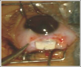
Fig. 2:   Postoperative picture showing scleral thinning strengthened by a scleral patch graft.


perforations similar to many other studies2,18. The biological quality of corneoscleral discs was reported comparable to that of tissue obtained from enucleated eye. Sclera (corneoscleral button) has number of advantages but the strict criticism was necrotic process. Similarly, peripheral corneal grafting is also the rare surgical treatments with tectonic sclera excluding in case of necrotizing sclera19. Sclera was also used as a graft in most of the studies, in scleromalacia. Similarly, there is a list of many tissues used as reconstructive materials but, still no such material is universally acceptable.


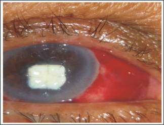
Fig. 3:   Severe corneal thinning with descemetocele covered with scleral patch graft.


In our study, males are more commonly affected than females similar to Shalini Mohan et.al. study in which five times more affected peoples are males than females15. The risk of damage was commonly found in young age group – around half of patients in our study were under 50 years of age. Detailed patient data which includes mode, duration and injury object are foremost step followed in any corneoscleral perforation repair. But, it was included in limitations of our study that no such related history was noted from patients. Patients before surgery were properly evaluated to the injury with other associated injuries for possibility of concomitant microbial contamination etc.

It is well known that surgical treatment alone does not solve the problem of the patient, therefore physician must control the immunoregulatory dysfunction which causes destruction of the graft and, subsequently, the patient's eye20.

After scleral graft visual acuity was improved in our study similar to study done by Hwan and coworkers21. Previous studies show that visual improvement was made by removing sutures on corneal side of scleral graft and by decreasing inflammation22. Ti et al, reported that after pterygium surgery in patients with scleral melting, corneal lamellar graft help to maintain integrity of the globe23. In this study, scleral patch grafting in spontaneous and traumatic corneoscleral perforations was achieved in most of the eyes for scleral defects of favorable structural outcome. Only, three patients had complications; two patients developed phithisical eye and one eye was eviscerated due to late onset endophthalmitis.

This study has numerous limitations, including the loss of patients to follow-up and incomplete records. Despite the numerous limitations, the study demonstrates the limitations of our current surgical capabilities to combat ocular trauma. Another limitation is the lack of details of re-epithelialization of the stable ocular surface.


CONCLUSION

This study concludes that preserved scleral graft in spontaneous and traumatic corneoscleral perforations provides functional and structural stability to eyes with rare complications.


Conflict of Interest

There is no conflict of interest.


Author’s Affiliation

Dr. Sharjeel Sultan

MBBS, DOMS, MCPS, FCPS, FRCS.

Assistant Professor Ophthalmology

Dow University of Health Sciences

Civil Hospital, Unit 2 Eye Department Karachi-Pakistan


Dr. Nisar A Siyal

MBBS, MCPS, FCPS

Assistant Professor Ophthalmology

Dow University of Health Sciences

Civil Hospital, Unit 2 Eye Department Karachi-Pakistan


Dr. Nargis Nizam Ashraf

MBBS, FCPS.

Assistant Professor Ophthalmology

Dow University of Health Sciences

Civil Hospital, Unit 2 Eye Department Karachi-Pakistan


Dr. A Rasheed Khokhar

MBBS, FCPS

Professor and Head of department ophthalmology

Dow University of Health Sciences

Civil Hospital, Unit 2 Eye Department Karachi-Pakistan

Role of Authors

Dr. Sharjeel Sultan

Concept and design, undertook the data analyses, wrote, edited and revised the manuscript.


Dr. Nisar A Siya

Interpretation of data, wrote and reviewed the manuscript


Dr. Nargis Nizam Ashraf

Wrote, edited and reviewed the manuscript


Dr. A Rasheed Khokhar

Reviewed and approved the manuscript


REFERENCES

1.        Jennifer HS, Marian S. M Needles, Sutures, and Instruments. Available from www.ophed.com

2.        Fong YY, Yu M, Young AL, Jhanji V. Presentation and management outcomes of corneal and scleral perforations in geriatric nursing home residents. Medicine, 2015; 94 (36).

3.        Shalini M, Anand A, Anita P, Shibal B. Repair of Corneoscleral Perforations. DOS Times– 2008; 14 (2).

4.        Dandona R, Dandona L. Corneal blindness in a southern Indian population: need for health promotion strategies. Br J Ophthalmol. 2003; 87 (2): 133-141.

5.        Sangwan VS, Jain V, Gupta P. Structural and functional outcome of scleral patch graft. Eye, 2007; 21 (7): 930-5.

6.        Nguyen QD, Foster CS. Scleral patch graft in the management of necrotizing scleritis. International ophthalmology clinics, 1999; 39 (1): 109-31.

7.        Parekh M, Ferrari S, Di Iorio E, Barbaro V, Camposampiero D, Karali M, Ponzin D, Salvalaio G. A simplified technique for in situ excision of cornea and evisceration of retinal tissue from human ocular globe. Journal of visualized experiments: JoVE. 2012; (64): 3765.

8.        Sangwan VS, Jain V, Gupta P. Structural and functional outcome of scleral patch graft. Eye, 2007; 21 (7): 930.

9.        Lee JS, Shin MK, Park JH, Park YM, Song M. Autologous advanced tenon grafting combined with conjunctival flap in scleromalacia after pterygium excision. Journal of ophthalmology, 2015; 2015.

10.     Ramenaden ER, Raiji VR. Clinical characteristics and visual outcomes in infectious scleritis: a review. Clinical Ophthalmology (Auckland, NZ). 2013; 7: 2113.

11.     Sahin I, Alhan D, Nışancı M, Ozer F, Eski M, Işık S. Auto-/homografting can work well even if both autograft and allograft are meshed in 4: 1 ratio. Ulusal travma ve acil cerrahi dergisi = Turkish journal of trauma & emergency surgery: TJTES. 2014; 20 (1): 33-8.

12.     Ti SE, Tan DT. Tectonic corneal lamellar grafting for severe scleral melting after pterygium surgery. Ophthalmology, 2003; 110 (6): 1126-36.

13.     Stunf, S., Lumi, X., & Drnovšek-Olup, B. Preserved scleral patch graft for unexpected extreme scleral thinning found at the scleral buckling procedure: A case report. Indian Journal of Ophthalmology, 2011; 59 (3): 235–238.

14.     Elisabeth, P., Hilde, B., & Ilse, C. Eye bank issues: II. Preservation techniques: warm versus cold storage. International Ophthalmology, 2008; 28 (3), 155–163.

15.     Yonekawa Y, Chodosh J, Eliott D. Surgical techniques in the management of perforating injuries of the globe. International ophthalmology clinics, 2013; 53 (4): 127-37.

16.     Vlasov A, Ryan DS, Ludlow S, Coggin A, Weichel ED, Stutzman RD, Bower KS, Colyer MH. Corneal and corneoscleral injury in combat ocular trauma from Operations Iraqi Freedom and Enduring Freedom. Military medicine, 2017; 182 (1): 114-9.

17.     Hamdi M, Hamdi I. Scleral Repair by Biodegradable Collagen Implant in Strabismus Surgery. Ophthalmol Res an Int J. 2015; 3 (4): 141-6.

18.     Repair of Corneoscleral Perforations (PDF Download Available). Available from:

https://www.researchgate.net/./234059015_Repair_of_Corneoscleral_Perforations.

19.     Nguyen QD, Foster CS. Scleral patch graft in the management of necrotizing scleritis. International ophthalmology clinics, 1999; 39 (1): 109-31.

20.     Parada-Vasquez RH, Benitez-Castrillon PC, de Leon-Ortega JE, Leon-Roldan CR. Scleral patch graft in the management of necrotizing scleritis with inflammation. A case report. Archivos de la Sociedad Espanola de Oftalmologia. 2016; 91 (7): 353.

21.     Hwan J, Kim JC. Repair of scleromalacia using preserved scleral graft with amniotic membrane transplantation. Cornea, 2003; 22: 288–293.

22.     Sangwan VS, Jain V, Gupta P. Structural and functional outcome of scleral patch graft. Eye, 2007; 21 (7): 930.

23.     Pai V, Shetty J, Amin H, Thomas M. Management of Scleral Thinning: An Alternate Approach. Nitte University Journal of Health Science, 2016; 6 (1): 88.